Proton beam therapy is a type of radiotherapy that can concentrate the dose on a target tumor by using the proton beam range based on the Bragg curve. The actual range differs depending on the internal structure of the region through which the beam passes; therefore, inaccurate patient positioning may cause deviations from the planned dose distribution. Accordingly, proton beam therapy requires patient positioning that takes the proton beam range and dose distribution into account.
Our laboratory focuses on cone-beam computed tomography (CBCT) scanning and reconstruction techniques as well as advanced image-guided technology using three-dimensional information about a patient’s body, which is obtained from CBCT images. More specifically, we study: dual-source four-dimensional CBCT (4D-CBCT) scanning and reconstruction techniques to generate clear three-dimensional images of a moving target based on X-ray fluoroscopic images acquired simultaneously from two directions; an image registration method using weighted normalized mutual information based on the size of dose delivered; and a positioning method with minimal dose errors through visualization and indexing of the penetration depth of proton beam (water-equivalent thickness) of each irradiation spot.
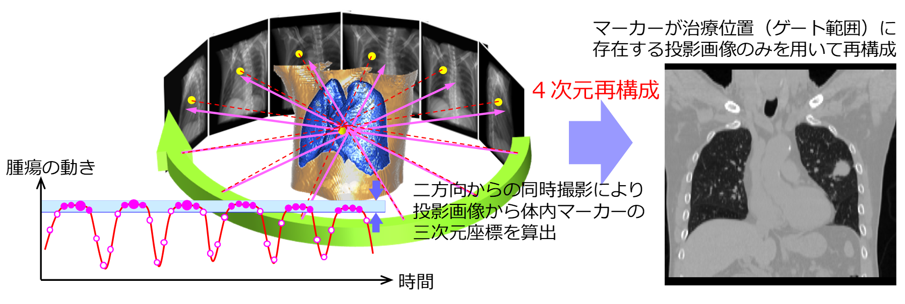
Principle of reconstruction of four-dimensional cone beam CT imaging
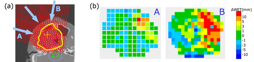
(a)Beam (spot) placement in head and neck cancer treatment planning (b)Differential water equivalent thickness map